Our Technology
About / Our Technology
Our Technology
At Buck and Todd Optometrists, we’re committed to staying at the forefront of technology. We continuously invest in cutting-edge equipment, pushing the boundaries of conventional vision care. Our expanding arsenal of advanced tools has revolutionised our ability to detect eye diseases early and take proactive measures. Whether it’s identifying glaucoma, macular degeneration, addressing dry eye, or managing complex contact lens fittings, no aspect of eye health escapes our meticulous attention. Coupled with our extensive experience, these cutting-edge tools empower us to deliver incomparable vision care for our patients.
Automated
Refractor

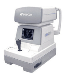
Automated Refractor
The automated refractor allows our optometrist to have a baseline for what your prescription may be based on the way light travels through your eye. The auto refractor does this by projecting an image in front of your eye, the light from this image passes through the cornea, pupil and lens of your eye and bounces off the retina (the back of your eye) and then reflects to a sensor in the refractor. This process creates a probable prescription. We have utilised this technology since 2006.
i-Care Tonometer

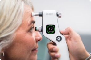
i-Care Tonometer
The i-Care tonometer is our new hand-held tool that measures the pressure in your eye. This offers a safe, quick, and easy way to measure the pressures and more comfortable for patients, no more puff of air! The i-Care tonometer simply taps the surface of the cornea 6 times and calculates an average pressure IOP measurement.
Corneal
Topography

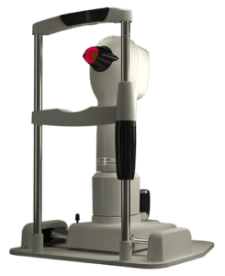
Corneal Topography
We have utilised the corneal topographer since 2006 to measure and assess the shape of the cornea. It is a quick and painless test that allows our optometrist to customise to fit of contact lenses to each individual eye. The topographer also allows us to do a tear film analysis. This shows us the structural and functional quality of your tear film and whether you need to be using lubrication drops etc.
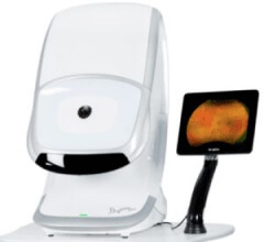
OPTOS
Daytona

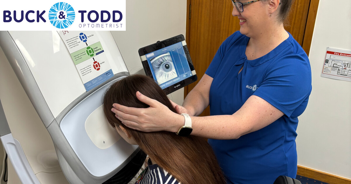
OPTOS Daytona
The OPTOS Daytona is a non-dilating camera which captures an anatomically correct, ultra-wide image of the retina in seconds. The scan is easy on the patient and gives the optometrist images instantly with advanced clarity. Unlike conventional devices, the OPTOS incorporates low-powered laser wavelengths rather than scanning simultaneously. The scans help our optometrists detect any abnormalities and diseases including glaucoma, diabetes, eye tumours, bleeding in the eye and retinal detachments.
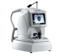
Ocular Coherence Tomography (OCT)

Ocular Coherence Tomography (OCT)
The ocular coherence tomography (RS-3000 RetinaScan) captures scans of your retina’s distinctive layers which allows our optometrists to map and measure your macular and retina. We are also able to measure and monitor the surface of the eye and thickness of the cornea. The OCT’s combination of high-speed and auto focus technology offers outstanding precision and ease-of-use. Capturing 53,000 A-scans a second helps to reduce the time taken to capture measurements and show the discrete retinal layers. Auto-tracking function monitors eye movement and any changes from previous visits. This all allows us to monitor glaucoma, macular degeneration, diabetic retinopathy, and retinal detachments closely as well as the general health of the eye.
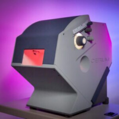
Intuitive
Colorimetry

Intuitive Colorimetry
Intuitive Colorimetry testing allows us to test visual stress and determine if coloured lenses are beneficial to improve the patient’s ability to read, use a computer etc. without headaches or eye strain. This technology projects different colours and shades onto a text and the patient compares colours to white light to see if any changes or comfort occurs. We are able to assess what hue and level of saturation of colour or shade is needed. We then take the results of this test and create a lens sample of the most comfort colour chosen. For more in-depth information on colorimetry click HERE.
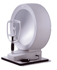
Visual
Fields Machine

Visual Fields Machine
The visual fields tests the quality of your peripheral vision in each eye individually and how much vision loss may be occurring overtime. This is done by placing the patient directly in line with a visual point and making that their central fixation. While the patient focuses on this point, lights will flash all around in their peripheral vision. As the patient successfully sees a flash, they click a button. The program connected to the machine calculates and records what is missed. These results then create a map of your vision which shows the optometrist if you may have blind spots in your vision and where they are occurring. The size and shape of these blind spots can determine if any eye diseases are affecting your vision like glaucoma, diabetes, high blood pressure and brain tumours.
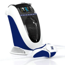
Intense Pulsed
Light (IPL)

Intense Pulsed Light (IPL)
IPL is a novel and extremely effective treatment for evaporative dry eye. IPL is a calibrated series of intense pulsating light which are set at a specific energy and frequent to stimulate the glands in your eyelids. This helps the glands recover function and loosen any clogged oils. After each treatment the optometrist will apply pressure to the glands to assess the colour and consistency of the oils. We take precautions when treatments patients with the light, applying protective patches to the eyes and lashes as well as gel to the skin. To read more about Dry eyes and our IPL therapy click HERE.
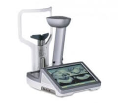
Lipiscan

Lipiscan
Our Lipiscan technology provides non-intrusive imaging of the eyelid glands. The images are captured using near infrared illumination from multiple light sources, reducing any reflections and creating a consistent light intensity across the surface of the eyelid. These scans expose and highlight the condition of the meibomian glands (the oil glands that line the edge of the eyelids). These scans play a role in diagnosing and treating symptoms of chronic dry eye.
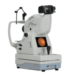
Retinal
Camera

Retinal Camera
Even though we have more advanced technology for imaging like our OPTOS and OCT we still like to use our Retinal camera to confirm any abnormalities or as an alternative for patients too young or unable to use our other equipment. Providing a 40-degree retinal picture in real colour the retinal camera helps evaluate for any artifacts in the 200-degree Optos scan.
