• Caring for the eyes of Mackay since 1952

Macular Degeneration

Macular Degeneration impairs a person’s ability to read, recognise faces, drive, and makes it difficult to recognise colours and contrasts.

Commonly affecting people over 50 years of age it is referred to as ‘age-related macular degeneration’ or AMD. There is currently no cure for AMD, and treatment efforts are directed at maintaining useful central vision for as long as possible.

It never causes complete blindness as the surrounding (side) vision remains normal, but it is the leading cause of vision loss in the western world, accounting for almost 50% of all visually impaired cases.

There are two types of AMD: dry (atrophic) and wet (neovascular or exudative). Most AMD starts as the dry type and in 10-20% of individuals, it progresses to the wet type. Age-related macular degeneration is always bilateral (i.e., occurs in both eyes), but does not necessarily progress at the same pace in both eyes. It is therefore possible to experience the wet type in one eye and the dry type in the other.

macula

Macular Degeneration is the leading cause of blindness in Australia and affects central vision.

Dry vs Wet Macular Degeneration

The dry (atrophic) type affects approximately 80-90% of individuals with AMD. It tends to progress more slowly than the wet type, and while there is not—as of yet—an approved treatment or cure most practitioners recommend a healthy diet, cessation of smoking and vitamin supplements to help maintain a healthier macula. In dry age-related macular degeneration, small white or yellowish deposits, called drusen, form beneath the macula causing it to deteriorate or degenerate over time.

The wet/neovascular type affects approximately 10-15% of individuals with age-related macular degeneration, but accounts for approximately 90% of all cases of severe vision loss from the disease.

In wet age-related macular degeneration, abnormal blood vessels under the retina begin to grow toward the macula. Because these new blood vessels are abnormal, they tend to break, bleed, and leak fluid, damaging the macula and causing it to lift up and pull away from its base. This can result in a rapid and severe loss of central vision.


Causes

The causes of AMD are not fully understood however the major risk factor is increasing age. The disease seems to cluster in certain families, suggesting that some are more genetically susceptible than others.


Symptoms

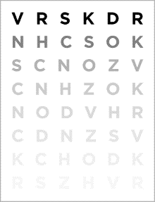
The main symptom of AMD is blurring of the central vision, with many people experiencing difficulty when reading. People also experience blurred, dark, or empty spots, similar to the after effect of looking into a flashbulb. A frequent and important symptom is distortion, straight lines such as door frames or street poles may appear bent or wavy.

Almost all patients with AMD can see well enough to take care of themselves and continue activities that do not require detailed vision.


Diagnosis

Regular eye examinations are the best way to detect AMD, this should be conducted by an Optometrist. If you experience any of the symptoms above, please make an appointment as soon as possible.


Risk Factors

Research suggests there are things that can be done to reduce the risk of developing AMD, including:

  • stop smoking,
  • eat a diet rich in fresh fruit and dark green leafy vegetables,
  • increase your intake of fish and other omega rich foods,
  • supplement your diet with vitamins, minerals and antioxidants,
  • protect your eyes from ultraviolet light,
  • check your vision regularly with your doctor.

Treatment

There is currently no cure for AMD, and treatment efforts are directed at maintaining useful central vision for as long as possible. Treatment varies depending on the type of AMD and individual characteristics of the condition.

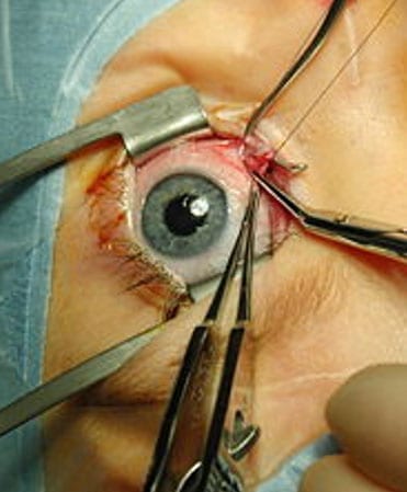
The most common treatment available for wet AMD is injection of Anti-VEGF agents: Ranibizumad (Lucentis), Aflibercept (Eylea) and Bevacizumab (Avastin). These drugs are injected into the vitreous cavity of the eye, reducing leakage from the blood vessels under the retina. Since this is not a cure, the majority of patients receiving these treatments will require life-long therapy at 1-3 monthly intervals.

There are currently no proven treatments to reverse the effects of dry AMD. Some vitamins, minerals and antioxidants ‘may’ slow down the progression of early stage of AMD

For more information see contact the Macular Disease Foundation: http://www.mdfoundation.com.au/

Dr Ieuan H. Rees. (Optometrist)
Buck & Todd Optometrist
Located at 103 Alfred Street, Mackay, 4740.

Mackay Optometrist | Mackay Eye Doctor Testing
Read More

Cataracts

Cataracts are opacification of the normally clear crystalline lens inside the eye. Our optometrist uses an internationally recognized classification system to allow tracking of progression of the opacity over time, whilst evaluating changes in visual acuity and contrast sensitivity.


As can be seen in in Lens Opacities Classification System image below, cataracts develop in different regions of the lens and vary in density and thus have varying effects on contrast and visual acuity. Some forms progress at faster rates than others, requiring more regular review.


Over time the clouded areas become larger and denser and cause the patient’s sight to become worse. The time taken for this to happen varies from a few months to many years. Usually cataracts affect both eyes, but often develop at different rates in each eye. The development of cataracts is a gradual painless worsening of sight. Other symptoms include blurred or hazy vision, spots before the eyes, double vision and a marked increase in sensitivity to glare.


An examination by our optometrist will reveal any changes that have occurred in the lens of the eye. We have special equipment that enables us to see changes in the lens that may lead to cataracts several years before symptoms appear. People older than 65 years often have signs of cataracts and should have their eyes examined regularly. If untreated, cataracts can cause blindness. Blindness can be prevented by detecting the cataracts early and, if necessary, by having them removed surgically.


Most patients have an intraocular lens (IOL) inserted at the time of surgery, with excellent results. This is a plastic lens that replaces your own cloudy lens. Patients may also need to wear spectacles or contact lenses.


Some surgeons now offer multi-focal, or extended focus intra-ocular lenses (MFIOL), allowing correction for both distance and near without glasses. These work to varying degrees of success with haloes around lights and decreased contrast sensitivity a common side effect. Generally, MFIOL work best for those who don’t drive much at night, are long-sighted, have regular corneal topography, healthy retinas, and have a relatively good tolerance to blur. Your suitability for a MFIOL should be discussed at length with our optometrists.


There is no proven method of preventing cataracts. Long term exposure to ultraviolet lights is thought to induce cataracts, so a brimmed hat and approved sunglasses should be worn in sunlight. Cataract surgery is performed when your vision interferes with daily life. We will assist you in making this decision. Cataract surgery is now a relatively minor procedure. Often it is performed under a local anesthetic on an out-patient basis. This means that the patient attends a hospital or clinic for the surgery and can go home the same day.


The surgery is performed by an ophthalmologist, a medical doctor who specializes in eye surgery. We will refer you to an ophthalmologist if necessary.

Dr Ieuan H. Rees. (Optometrist)
Buck & Todd Optometrist
Located at 103 Alfred Street, Mackay, 4740.

Mackay Optometrist | Mackay Eye Doctor Testing
Read More

Myopia = Short-sightedness

Myopia typically begins in childhood and tends to progress; with stronger prescription glasses required each year. In most cases, myopia stabilizes around age 25 years but sometimes it continues to progress with age.

If you are myopic or near-sighted, you typically will have difficulty reading road signs, the board at school and seeing distant objects clearly. Generally you see well for close-up tasks such as reading and computer use. Myopia occurs when the eyeball is too long, relative to the combined focusing power of the cornea and lens. This causes light rays to focus at a point in front of the retina, rather than directly on its surface.

Prevalence of myopia in children and young adults is increasing. Earlier age of onset is linked to faster myopic progression, which in turn contributes to the health implications of progressive myopia that place children at risk for eye diseases. The lifetime risks of myopic pathologies such as myopic maculopathy, cataract, glaucoma and retinal detachment are significant. Myopia is a pathological condition and there are no “safe” levels.

We can slow myopia: research shows there are a number of ways we can slow myopia progression in children, and in turn reduce risk of eye disease. “Whilst there is no magic cure for myopia control, the child at-risk of developing myopia, should not have to suffer from inaction:-Inaction is no longer acceptable.”

We can slow and even stop myopia progression. Research shows there are a number of ways we can slow myopia progression in children. Altering optics, pharmacological agents and behavioural modifications, individually, or in tandem have proven to be effective to varying and cumulative degrees.

Pharmacological agents

Daily low dose 0.02-0.05% atropine is reasonably effective (64%) in slowing myopia progression. However even at this minimal concentration, glare sensitivity and need for reading glasses can occur. “WARNING” Long term use of anticholinergic medications has recently been linked to cognitive impairment and earlier onset of dementia.

To determine your child’s myopia risk profile please review the following chart. Our optometrist can guide you through this profile and give advice on what you can do to mitigate the risk

Please click the image below for a full size chart

Outdoor Activity

Outdoor activity reduces risk of myopia development in pre-teenage children. 2 hours per day outside daylight activity is recommended. Please remember to slip, slop, slap and slide on some sunglasses.

Reading Glasses

Multifocal, bifocal, extended focus and reading glasses help control any residual over-convergence and reduce the need for excess accommodation. These slow myopia (25% effective) only when there is over-convergence at near. Plus powered readers will often prove helpful as an adjunctive therapy. Full correction and/or under-correction single vision glasses do little to slow myopia progression. Myopia inevitably progresses and stronger prescription lenses are required each year.

Myovision Spectacles

The desired change in optics required to reduce myopic progression is difficult to achieve with spectacle lenses. Lenses such as Myovision, designed to slow progression of myopia have shown variable results. Inability to keep optical control on visual axis being the major limitation.

Contact Lenses

Negatively powered contact lenses (0% effective) induce negative spherical aberration placing the focus slightly behind the retina, increasing accommodative demand and reducing focus accuracy. This augments the effects of peripheral defocus behind the retina and results in the signal for increased axial eye growth.

Distance centred multifocal soft contact lenses have shown myopia control results ranging from 29% to 50% effectiveness over 24 months. The optics create a similar optical effect to orthokeratology but have yet to prove to be as effective.

Vision Therapy

There is an association between higher levels of over-convergence and reduced focussing reserve at near in myopic children along with greater variability in accommodative responses. Vision therapy improves binocular co-ordination, peripheral awareness, reduces accommodative stress and strengthens vergence reserves. Re-organising the visual environment, reduces focussing stress and helps slow myopia progression.

Orthokeratology

Orthokeratology involves custom designed contact lens retainers which are worn during sleep. Similar to retainers for the teeth, these lenses adjust the shape of the cornea, correcting for myopia by flattening the central cornea whilst steepening the mid-peripheral cornea. This provides clear vision all day, without glasses or contact lenses and shifts the peripheral optics of the myopic eye from behind, to in front of the retina, whilst pushing the central focus back onto the fovea providing clear vision. This change in the shape of the image shell slows axial elongation and is 85% effective as a stand-alone treatment in stopping myopia progression.

Adjusting the mid-peripheral optics helps counter accommodation errors, improves functional peripheral visual fields, reduces esophoria and aids focussing all whilst reducing the signal for myopia progression. 20/20 unaided vision can be restored in as little as two days to one week depending on severity.

Dual/combined therapy such as reading glasses, atropine and vision therapy can be used in combination with orthokeratology to gain greater myopia control.

For more information about Orthokeratology please visit the following websites:

www.orthokdoctors.com

www.myopiaprevention.org/references_orthokeratology.html

NaturalVue and MiVision

Two recently designed contact lenses based on the optics of orthokeratology vision results are NaturalVue and MiVision. These provide similar peripheral optics results to orthokeratology, for myopes, when worn on a daily wear basis. The effect is only effective when the lenses are worn and thus lack the 24/7 effect of orthokeratology. Reported success rates in slowing/stopping myopia progression are around 65%.

Dr Ieuan H. Rees. (Optometrist)
Buck & Todd Optometrist
Located at 103 Alfred Street, Mackay, 4740.

Mackay Optometrist | Mackay Eye Doctor Testing
Read More

Low Vision

Low vision describes the level of vision between being fully sighted and totally blind. Having low vision upsets a person’s ability to carry out many of the daily tasks for which vision is essential. Fortunately, the remaining vision can often be used effectively with the help of magnifiers and other vision aides. Many optometrists specialise in helping people with moderate and severe vision impairment. They can help patients make the best use of their remaining vision, even when they have been told that nothing more can be done to treat the medical cause of vision impairment.

Q: What is low vision?

Low vision is clinically defined as a specific range of visual acuity, which is the ability to read letters on an eye testing chart of decreasing size.

Other factors affecting low vision are a person’s ability to detect objects in their side or peripheral vision. And their sensitivity to medium and low contrast.

All of these functions may be impaired in some conditions that cause low vision. Practical definition state that a person has low vision when, as a result of eye conditions or disease, he or she cannot perform vision tasks that ordinarily are not difficult, or they do not meet the vision standards for driving.

Q: What causes low vision?

No age is exempt from low vision. Infants and children can be born with inherited developmental anomalies of the eye that cause low vision. About 90 per cent of children with low vision have a stable ocular condition so their vision is unlikely to deteriorate in working adulthood. Low vision care for these children offers life-long benefits.

Young adults might suffer from inherited conditions that manifest later in life. A loss of central vision occurs in macular dystrophies such as Stargardt’s disease, while peripheral vision loss is progressive in retinitis pigmentosa. General health conditions such as diabetes can progress to include retinal complications and cataracts.

Low vision is far more common in older people.

Macular degeneration, diabetes and glaucoma are common conditions that are sometimes difficult or impossible to cure with medical treatment.

Medical condition such as strokes and brain tumours can rob a person of half their visual field. Optometrist provide advice on magnifiers, managing visual field losses, use of lighting, access to low vision services and lifestyle changes to make living with low vision more tolerable. Referral for specialist care is also available.

Dr Ieuan H. Rees. (Optometrist)
Buck & Todd Optometrist
Located at 103 Alfred Street, Mackay, 4740.

Mackay Optometrist | Mackay Eye Doctor Testing
Read More

Laser Refractive Surgery

(LASIK, PRK, SMILE) 

Surgical procedures aimed at reducing the refractive error of the eye are called refractive surgery. In LASIK, PRK and SMILE, precise and controlled removal of corneal tissue by a special laser reshapes the cornea changing its focusing power.

Radial Keratotomy or RK developed in Russia has been long surpassed by PRK (Photorefractive Keratectomy) LASIK and now SMILE. PRK was the first surgical procedure developed to reshape the cornea, by sculpting, using a laser. Often the exact same laser is used but the way the stroma (the middle layer of the cornea), is vaporized by the laser vaies. In PRK, the top layer of the cornea, called the epithelium, is scraped away to expose the stromal layer underneath. In LASIK, a flap is cut in the stromal layer and the flap is folded back. In SMILE the laser creates a lenticule inside the stroma which is then extracted.

When is LASIK not for me?

You are probably NOT a good candidate for refractive surgery if:

  • You are not a risk taker. Certain complications are unavoidable in a percentage of patients, and there are no long-term data available for current procedures.
  • It will jeopardize your career. Some jobs prohibit certain refractive procedures. Be sure to check with your employer/professional society/military service before undergoing any procedure.
  • Cost is an issue. Most medical insurance will not pay for refractive surgery. Although the cost is coming down, it is still significant.
  • You required a change in your contact lens or glasses prescription in the past year. This is called refractive instability.
    Patients who are more likely to have refractive instability should discuss the possible additional risks with our optometrists, particularly those:
  • In their early 20s or younger,
  • Whose hormones are fluctuating due to disease such as diabetes,
  • Who are pregnant or breastfeeding, or
  • Who are taking medications that may cause fluctuations in vision,
  • You have a disease or are on medications that may affect wound healing. Certain conditions, such as autoimmune diseases (e.g., lupus, rheumatoid arthritis), immunodeficiency states (e.g., HIV) and diabetes, and some medications (e.g., retinoic acid and steroids) may prevent proper healing after a refractive procedure.
  • You actively participate in contact sports. You participate in boxing, wrestling, martial arts or other activities in which blows to the face and eyes are a normal occurrence.
  • You are not an adult.  Currently, no lasers are approved for LASIK on persons under the age of 18.

Precautions
The safety and effectiveness of refractive procedures has not been determined in patients with some diseases. Discuss with your doctor if you have a history of any of the following:

  • Herpes simplex or Herpes zoster (shingles) involving the eye area.
  • Glaucoma, glaucoma suspect, or ocular hypertension.
  • Eye diseases, such as uveitis/iritis (inflammations of the eye)
  • Eye injuries or previous eye surgeries.
  • Keratoconus

Other Risk Factors
Your doctor should screen you for the following conditions or indicators of risk:

  • Blepharitis. Inflammation of the eyelids with crusting of the eyelashes, that may increase the risk of infection or inflammation of the cornea after LASIK.
  • Large pupils. Make sure this evaluation is done in a dark room. Although anyone may have large pupils, younger patients and patients on certain medications may be particularly prone to having large pupils under dim lighting conditions. This can cause symptoms such as glare, halos, starbursts, and ghost images (double vision) after surgery. In some patients these symptoms may be debilitating. For example, a patient may no longer be able to drive a car at night or in certain weather conditions, such as fog.
  • Thin Corneas. The cornea is the thin clear covering of the eye that is over the iris, the coloured part of the eye. Most refractive procedures change the eye’s focusing power by reshaping the cornea (for example, by removing tissue).  Performing a refractive procedure on a cornea that is too thin may result in blinding complications.
  • Previous refractive surgery (e.g., RK, PRK, LASIK).  Additional refractive surgery may not be recommended.  The decision to have additional refractive surgery must be made in consultation with your doctor after careful consideration of your unique situation.
  • Dry Eyes. LASIK surgery tends to aggravate this condition.

What are the risks and how can I find the right doctor for me?

Most patients are very pleased with the results of their refractive surgery. However, like any other medical procedure, there are risks involved. That’s why it is important for you to understand the limitations and possible complications of refractive surgery.

Before undergoing a refractive procedure, you should carefully weigh the risks and benefits based on your own personal value system, and try to avoid being influenced by friends that have had the procedure or doctors encouraging you to do so.

  • Some patients lose vision. Some patients lose lines of vision on the vision chart that cannot be corrected with glasses, contact lenses, or surgery as a result of treatment.
  • Some patients develop debilitating visual symptoms. Some patients develop glare, halos, and/or double vision that can seriously affect night-time vision. Even with good vision on the vision chart, some patients do not see as well in situations of low contrast, such as at night or in fog, after treatment as compared to before treatment.
  • You may be under treated or over treated. Only a certain percent of patients achieve 20/20 vision without glasses or contacts. You may require additional treatment, but additional treatment may not be possible. You may still need glasses or contact lenses after surgery. This may be true even if you only required a very weak prescription before surgery. If you used reading glasses before surgery, you may still need reading glasses after surgery.
  • Some patients may develop severe dry eye syndrome. As a result of surgery, your eye may not be able to produce enough tears to keep the eye moist and comfortable. Dry eye not only causes discomfort but can reduce visual quality due to intermittent blurring and other visual symptoms. This condition may be permanent. Intensive drop therapy and use of plugs or other procedures may be required.
  • Results are generally not as good in patients with very large refractive errors of any type. You should discuss your expectations with your doctor and realize that you may still require glasses or contacts after the surgery.
  • For some farsighted patients, results may diminish with age. If you are farsighted, the level of improved vision you experience after surgery may decrease with age. This can occur if your manifest refraction (a vision exam with lenses before dilating drops) is very different from your cycloplegic refraction (a vision exam with lenses after dilating drops).
  • Long-term data are not available. LASIK is a relatively new technology. The first laser was approved for LASIK eye surgery in 1998. Therefore, the long-term safety and effectiveness of LASIK surgery is not known.

Additional Risks if you are Considering the Following:

  • Monovision

Monovision is one clinical technique used to deal with the correction of presbyopia, the gradual loss of the ability of the eye to change focus for close-up tasks that progresses with age. The intent of monovision is for the presbyopic patient to use one eye for distance viewing and one eye for near viewing. This practice was first applied to fit contact lens wearers and more recently to LASIK and other refractive surgeries. With contact lenses, a presbyopic patient has one eye fit with a contact lens to correct distance vision, and the other eye fit with a contact lens to correct near vision. In the same way, with LASIK, a presbyopic patient has one eye operated on to correct the distance vision, and the other operated on to correct the near vision. In other words, the goal of the surgery is for one eye to have vision worse than 20/20, the commonly referred to goal for LASIK surgical correction of distance vision. Since one eye is corrected for distance viewing and the other eye is corrected for near viewing, the two eyes no longer work together. This results in poorer quality vision and a decrease in depth perception. These effects of monovision are most noticeable in low lighting conditions and when performing tasks requiring very sharp vision. Therefore, you may need to wear glasses or contact lenses to fully correct both eyes for distance or near when performing visually demanding tasks, such as driving at night, operating dangerous equipment, or performing occupational tasks requiring very sharp close vision (e.g., reading small print for long periods of time).

Many patients cannot get used to having one eye blurred at all times. Therefore, if you are considering monovision with LASIK, make sure you go through a trial period with contact lenses to see if you can tolerate monovision, before having the surgery performed on your eyes. Find out if you pass your state’s driver’s license requirements with monovision.

In addition, you should consider how much your presbyopia is expected to increase in the future. Ask your doctor when you should expect the results of your monovision surgery to no longer be enough for you to see near-by objects clearly without the aid of glasses or contacts, or when a second surgery might be required to further correct your near vision.

  • Bilateral Simultaneous Treatment

You may choose to have LASIK surgery on both eyes at the same time or to have surgery on one eye at a time. Although the convenience of having surgery on both eyes on the same day is attractive, this practice is riskier than having two separate surgeries.

If you decide to have one eye done at a time, you and your doctor will decide how long to wait before having surgery on the other eye. If both eyes are treated at the same time or before one eye has a chance to fully heal, you and your doctor do not have the advantage of being able to see how the first eye responds to surgery before the second eye is treated.

Another disadvantage to having surgery on both eyes at the same time is that the vision in both eyes may be blurred after surgery until the initial healing process is over, rather than being able to rely on clear vision in at least one eye at all times.

Finding the Right Doctor
If you are considering refractive surgery, make sure you:

  • Compare. The levels of risk and benefit vary slightly not only from procedure to procedure, but from device to device depending on the manufacturer, and from surgeon to surgeon depending on their level of experience with a particular procedure.
  • Don’t base your decision simply on cost and don’t settle for the first eye center, doctor, or procedure you investigate. Remember that the decisions you make about your eyes and refractive surgery will affect you for the rest of your life.
  • Be wary of eye centers that advertise, “20/20 vision or your money back” or “package deals.” There are never any guarantees in medicine.
  • Read. It is important for you to read the patient handbook provided to your doctor by the manufacturer of the device used to perform the refractive procedure. Your doctor should provide you with this handbook and be willing to discuss his/her outcomes (successes as well as complications) compared to the results of studies outlined in the handbook.

Even the best screened patients under the care of most skilled surgeons can experience serious complications.

  • During surgery. Malfunction of a device or other error, such as cutting a flap of cornea through and through instead of making a hinge during LASIK surgery, may lead to discontinuation of the procedure or irreversible damage to the eye.
  • After surgery. Some complications, such as migration of the flap, inflammation or infection, may require another procedure and/or intensive treatment with drops. Even with aggressive therapy, such complications may lead to temporary loss of vision or even irreversible blindness.

Under the care of an experienced doctor, carefully screened candidates with reasonable expectations and a clear understanding of the risks and alternatives are likely to be happy with the results of their refractive procedure.

Advertising
Be cautious about “slick” advertising and/or deals that sound “too good to be true.” Remember, they usually are. There is a lot of competition resulting in a great deal of advertising and bidding for your business. Do your homework.

If you want to know more about advertising ethics, do’s and don’ts, or want to report on false advertising, explore the nearby Useful Links.

What should I expect before, during, and after surgery?

What to expect before, during, and after surgery will vary from doctor to doctor and patient to patient.  This section is a compilation of patient information developed by manufacturers and healthcare professionals, but cannot replace the dialogue you should have with your doctor.  Read this information carefully and with the checklist, discuss your expectations with your doctor.

Before Surgery

If you decide to go ahead with LASIK surgery, you will need an initial or baseline evaluation by your eye doctor to determine if you are a good candidate. This is what you need to know to prepare for the exam and what you should expect:

If you wear contact lenses, it is a good idea to stop wearing them before your baseline evaluation and switch to wearing your glasses full-time. Contact lenses change the shape of your cornea for up to several weeks after you have stopped using them depending on the type of contact lenses you wear. Not leaving your contact lenses out long enough for your cornea to assume its natural shape before surgery can have negative consequences. These consequences include inaccurate measurements and a poor surgical plan, resulting in poor vision after surgery. These measurements, which determine how much corneal tissue to remove, may need to be repeated at least a week after your initial evaluation and before surgery to make sure they have not changed, especially if you wear RGP or hard lenses.  If you wear:

  • soft contact lenses, you should stop wearing them for 2 weeks before your initial evaluation.
  • toric soft lenses or rigid gas permeable (RGP) lenses, you should stop wearing them for at least 3 weeks before your initial evaluation.
  • hard lenses, you should stop wearing them for at least 4 weeks before your initial evaluation.

You should tell your doctor:

  • about your past and present medical and eye conditions
  • about all the medications you are taking, including over-the-counter medications and any medications you may be allergic to

Your doctor should perform a thorough eye exam and discuss:

  • whether you are a good candidate
  • what the risks, benefits, and alternatives of the surgery are
  • what you should expect before, during, and after surgery
  • what your responsibilities will be before, during, and after surgery

You should have the opportunity to ask your doctor questions during this discussion.  Give yourself plenty of time to think about the risk/benefit discussion, to review any informational literature provided by your doctor, and to have any additional questions answered by your doctor before deciding to go through with surgery and before signing the informed consent form.

You should not feel pressured by your doctor, family, friends, or anyone else to make a decision about having surgery. Carefully consider the pros and cons.

The day before surgery, you should stop using:

  • creams
  • lotions
  • makeup
  • perfumes

These products as well as debris along the eyelashes may increase the risk of infection during and after surgery. Your doctor may ask you to scrub your eyelashes for a period of time before surgery to get rid of residues and debris along the lashes.

Also before surgery, arrange for transportation to and from your surgery and your first follow-up visit. On the day of surgery, your doctor may give you some medicine to make you relax. Because this medicine impairs your ability to drive and because your vision may be blurry, even if you don’t drive make sure someone can bring you home after surgery.

During Surgery

The surgery should take less than 30 minutes. You will lie on your back in a reclining chair in an exam room containing the laser system. The laser system includes a large machine with a microscope attached to it and a computer screen. (See the animation of the LASIK procedure.)

A numbing drop will be placed in your eye, the area around your eye will be cleaned, and an instrument called a lid speculum will be used to hold your eyelids open.

Your doctor may use a mechanical microkeratome (a blade device) to cut a flap in the cornea.

If a mechanical microkeratome is used, a ring will be placed on your eye and very high pressures will be applied to create suction to the cornea. Your vision will dim while the suction ring is on and you may feel the pressure and experience some discomfort during this part of the procedure. The microkeratome, a cutting instrument, is attached to the suction ring. Your doctor will use the blade of the microkeratome to cut a flap in your cornea. Microkeratome blades are meant to be used only once and then thrown out. The microkeratome and the suction ring are then removed.

Your doctor may use a laser keratome (a laser device), instead of a mechanical microkeratome, to cut a flap on the cornea.

If a laser keratome is used, the cornea is flattened with a clear plastic plate. Your vision will dim and you may feel the pressure and experience some discomfort during this part of the procedure. Laser energy is focused inside the cornea tissue, creating thousands of small bubbles of gas and water that expand and connect to separate the tissue underneath the cornea surface, creating a flap. The plate is then removed.

You will be able to see, but you will experience fluctuating degrees of blurred vision during the rest of the procedure. The doctor will then lift the flap and fold it back on its hinge, and dry the exposed tissue.

The laser will be positioned over your eye and you will be asked to stare at a light. This is not the laser used to remove tissue from the cornea. This light is to help you keep your eye fixed on one spot once the laser comes on. NOTE: If you cannot stare at a fixed object for at least 60 seconds, you may not be a good candidate for this surgery.

When your eye is in the correct position, your doctor will start the laser. At this point in the surgery, you may become aware of new sounds and smells. The pulse of the laser makes a ticking sound. As the laser removes corneal tissue, some people have reported a smell similar to burning hair. A computer controls the amount of laser energy delivered to your eye. Before the start of surgery, your doctor will have programmed the computer to vaporize a particular amount of tissue based on the measurements taken at your initial evaluation. After the pulses of laser energy vaporize the corneal tissue, the flap is put back into position.

A shield should be placed over your eye at the end of the procedure as protection, since no stitches are used to hold the flap in place. It is important for you to wear this shield to prevent you from rubbing your eye and putting pressure on your eye while you sleep, and to protect your eye from accidentally being hit or poked until the flap has healed.

After Surgery

Immediately after the procedure, your eye may burn, itch, or feel like there is something in it. You may experience some discomfort, or in some cases, mild pain and your doctor may suggest you take a mild pain reliever. Both your eyes may tear or water. Your vision will probably be hazy or blurry. You will instinctively want to rub your eye, but don’t! Rubbing your eye could dislodge the flap, requiring further treatment. In addition, you may experience sensitivity to light, glare, starbursts or haloes around lights, or the whites of your eye may look red or bloodshot. These symptoms should improve considerably within the first few days after surgery. You should plan on taking a few days off from work until these symptoms subside. You should contact your doctor immediately and not wait for your scheduled visit, if you experience severe pain, or if your vision or other symptoms get worse instead of better.

You should see your doctor within the first 24 to 48 hours after surgery and at regular intervals after that for at least the first six months. At the first postoperative visit, your doctor will remove the eye shield, test your vision, and examine your eye. Your doctor may give you one or more types of eye drops to take at home to help prevent infection and/or inflammation. You may also be advised to use artificial tears to help lubricate the eye. Do not resume wearing a contact lens in the operated eye, even if your vision is blurry.

You should wait one to three days following surgery before beginning any non-contact sports, depending on the amount of activity required, how you feel, and your doctor’s instructions.

To help prevent infection, you may need to wait for up to two weeks after surgery or until your doctor advises you otherwise before using lotions, creams, or make-up around the eye. Your doctor may advise you to continue scrubbing your eyelashes for a period of time after surgery. You should also avoid swimming and using hot tubs or whirlpools for 1-2 months.

Strenuous contact sports such as boxing, football, karate, etc. should not be attempted for at least four weeks after surgery. It is important to protect your eyes from anything that might get in them and from being hit or bumped.

During the first few months after surgery, your vision may fluctuate.

  • It may take up to three to six months for your vision to stabilize after surgery.
  • Glare, haloes, difficulty driving at night, and other visual symptoms may also persist during this stabilization period. If further correction or enhancement is necessary, you should wait until your eye measurements are consistent for two consecutive visits at least 3 months apart before re-operation.
  • It is important to realize that although distance vision may improve after re-operation, it is unlikely that other visual symptoms such as glare or haloes will improve.
  • It is also important to note that no laser company has presented enough evidence for the FDA to make conclusions about the safety or effectiveness of enhancement surgery.

Contact your eye doctor immediately, if you develop any new, unusual or worsening symptoms at any point after surgery. Such symptoms could signal a problem that, if not treated early enough, may lead to a loss of vision.

LASIK Surgery Checklist

Know what makes you a poor candidate

  • Career impact – does your job prohibit refractive surgery?
  • Cost – can you really afford this procedure?
  • Medical conditions – e.g., do you have an autoimmune disease or other major illness? Do you have a chronic illness that might slow or alter healing?
  • Eye conditions – do you have or have you ever had any problems with your eyes other than needing glasses or contacts?
  • Medications – do you take steroids or other drugs that might prevent healing?
  • Stable refraction – has your prescription changed in the last year?
  • High or Low refractive error – do you use glasses/contacts only some of the time? Do you need an unusually strong prescription?
  • Pupil size – are your pupils extra large in dim conditions?
  • Corneal thickness – do you have thin corneas?
  • Tear production – do you have dry eyes?

Know all the risks and procedure limitations

  • Over-treatment or under-treatment – are you willing and able to have more than one surgery to get the desired result?
  • May still need reading glasses – do you have presbyopia?
  • Results may not be lasting – do you think this is the last correction you will ever need? Do you realize that long-term results are not known?
  • May permanently lose vision – do you know some patients may lose some vision or experience blindness?
  • Dry eyes – do you know that if you have dry eyes they could become worse, or if you don’t have dry eyes before you could develop chronic dry eyes as a result of surgery?
  • Development of visual symptoms – do you know about glare, halos, starbursts, etc. and that night driving might be difficult?
  • Contrast sensitivity – do you know your vision could be significantly reduced in dim light conditions?
  • Bilateral treatment – do you know the additional risks of having both eyes treated at the same time?
  • Patient information – have you read the patient information booklet about the laser being used for your procedure?

Know how to find the right doctor

  • Experienced – how many eyes has your doctor performed LASIK surgery on with the same laser?
  • Equipment – does your doctor use an FDA-approved laser for the procedure you need? Does your doctor use each microkeratome blade only once?
  • Informative – is your doctor willing to spend the time to answer all your questions?
  • Long-term care – does your doctor encourage follow-up and management of you as a patient?  Your preop and postop care may be provided by a doctor other than the surgeon.
  • Be comfortable – do you feel you know your doctor and are comfortable with an equal exchange of information?

Know preoperative, operative, and postoperative expectations

  • No contact lenses prior to evaluation and surgery – can you go for an extended period of time without wearing contact lenses?
  • Have a thorough exam – have you arranged not to drive or work after the exam?
  • Read and understand the informed consent – has your doctor given you an informed consent form to take home and answered all your questions?
  • No makeup before surgery – can you go 24-36 hours without makeup prior to surgery?
  • Arrange for transportation – can someone drive you home after surgery?
  • Plan to take a few days to recover – can you take time off to take it easy for a couple of days if necessary?
  • Expect not to see clearly for a few days – do you know you will not see clearly immediately?
  • Know sights, smells, sounds of surgery – has your doctor made you feel comfortable with the actual steps of the procedure?
  • Be prepared to take drops/medications – are you willing and able to put drops in your eyes at regular intervals?
  • Be prepared to wear an eye shield – do you know you need to protect the eye for a period of time after surgery to avoid injury?
  • Expect some pain/discomfort – do you know how much pain to expect?
  • Know when to seek help – do you understand what problems could occur and when to seek medical intervention?
  • Know when to expect your vision to stop changing – are you aware that final results could take months?
  • Make sure your refraction is stable before any further surgery – if you don’t get the desired result, do you know not to have an enhancement until the prescription stops changing?

Other Resources

Below are some resources you may find helpful

Dr Ieuan H. Rees. (Optometrist)
Buck & Todd Optometrist
Located at 103 Alfred Street, Mackay, 4740.

Mackay Optometrist | Mackay Eye Doctor Testing
Read More

Multifocal Lenses

Multifocal lenses are required due to presbyopia (aging eye). They enable the wearer to focus on both near and distance and, depending on the lens type, middle distances, without removing his or her spectacles.

There are three main kinds of multifocal lenses: bifocal, trifocal and progressive.

Presbyopia

The crystalline lens inside the human eye changes its shape to let us see clearly at different distances. As we get older the lens begins to lose its flexibility, which makes it increasingly more difficult and tiring for us to change our focus and to see close objects clearly. The difficulty in focusing generally becomes noticeable about the age of 40 to 45 years. Presbyopia can be corrected by spectacle lenses that restore our power to see at near range.

Reading spectacles are prescribed to help people see at near. These spectacles cannot be used to see in the distance, so the wearer will continually find it necessary to remove the reading spectacles when looking at people, viewing television or looking across the room or the street. While driving, it is necessary to constantly scan the road and the instrument panel and a single-vision reading lens will not permit this.

Bifocal lenses

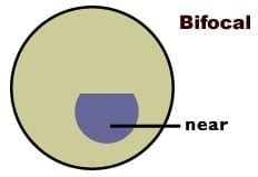
There are two distinct areas to a bifocal lens. In most cases the top portion is used for distance viewing and the lower portion for reading and other close work such as sewing.

The lower portion may be semi-circular, D-shaped, a narrow band or simply divided from the upper portion by a straight line. We can help you choose the kind of bifocal that will best suit your requirements.

If spectacles are already worn for distance vision, the onset of presbyopia means you will need either a second pair of spectacles for reading range or bifocal lenses incorporating prescriptions for both distance and near ranges.

For people who need near-range correction only, bifocals are still an excellent alternative to single vision lenses because they will avoid the inconvenience of removing the spectacles for distance vision.

Trifocal lenses

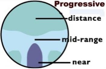
It is important for some people to be able to focus clearly at mid-range between distance and near. For example, Musicians may need to see their instruments close-up and read music at arm’s length while still being able to see the conductor in the distance. Someone with these needs will require trifocals, which give clear vision at three distances- far distance, middle distance and near.

There are three portions to a trifocal lens. The top, which is usually the largest portion, is for distance; the middle section is for seeing at intermediate distances such as at arm’s length; and the lowest part is for reading and other close work.

The dividing lines between the three portions may be curved or straight, just as with bifocals. Trifocal lenses have these portions in different positions to cater for your particular needs.

Progressive lenses

In progressive lenses, or varifocals as they are also called, the lens power changes gradually from distance correction through the intermediate powers to the reading prescription.

Unlike a trifocal, progressive lenses do not have definite dividing lines between the different portions of the lens. Instead of distinct segments, there is a gentle change from distant to intermediate to near focus parts of the lens.

The wearer has an infinite number of focusing distances; the nearest one can get to having natural sight with prescription spectacles. All distances are sharp and clear.

Most people think that the progressive lenses look better than bifocals or trifocals because, like ordinary single vision lenses, there are no obvious lines across the lenses. Some people are reluctant to wear multifocal lenses because they associate them with older people. Progressives offer them the best of both worlds- the benefits of Multifocal lenses and a young appearance.

Another advantage of progressive lenses is that because there isn’t any sharp line dividing portions of the lens, there is no obstruction to vision and no image jump, which makes it safer to use stairs and escalators.

Adapting to progressive lenses often takes a few weeks. Distortion to the sides of the lenses when first wearing progressives is a common finding. Most people do adapt as with any new pair of glasses, but frame adjustment and fit, as well as accurate measurement of visual axes are critical to success with progressive lens wear.

Just as people’s circumstances are different, so their spectacle requirements are different. What is best for one may not suit another. Your optometrist will be happy to discuss your requirements with you and advise you on the most appropriate lens type for your lifestyle. You may find you require different lenses for different purposes. Progressive lens designs which suit office work, may not suit driving or recreation such as golf.

Related Information.

  • Presbyopia
  • Bifocals

Dr Ieuan H. Rees. (Optometrist)
Buck & Todd Optometrist
Located at 103 Alfred Street, Mackay, 4740.

Mackay Optometrist | Mackay Eye Doctor Testing
Read More

Keratoconus

Keratoconus (literally, conical cornea) is a thinning and steepening of the central zone of the cornea, the front surface of the eye. These ‘wonky windows’ cause irregular astigmatism, where the unusual shape of the cornea causes the light to bend as it passes through, resulting in blurred vision. If left untreated, keratoconus can lead to severe vision impairment.

An inherited disorder, keratoconus occurs in about 1 in 800 people. Considered a recessive condition it requires genetic factors to be inherited from both parents. It is exacerbated by eye rubbing. Keratoconus usually becomes apparent between the ages of 10 and 25 years. It is sometimes associated with other conditions such as allergies, infantile eczema, asthma, reduced night vision, double jointed-ness and, in rare instances, with occasional short bouts of chest pain.

Glasses and contact lenses can give good vision and collagen cross linking surgery can be used to slow progression and penetrating keratoplasty may be required to treat severe cases. Collagen crosslinking, which increases the stiffness of the cornea, has been introduced as a treatment with reported success in slowing progression. However, the procedure is only suitable for keratoconus patients at the early stages of disease, and long-term outcomes are not yet known.

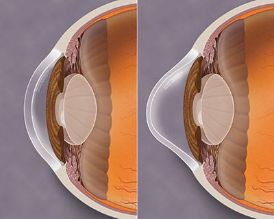
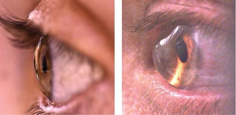
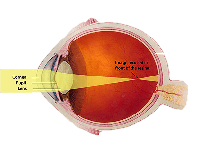
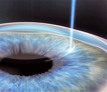
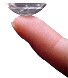
The initial symptom of keratoconus is blurred vision, which is caused by shortsightedness and astigmatism. At this stage, good vision generally can be obtained with spectacles. As keratoconus progresses, the shape of the cornea becomes irregular and it is not possible to correct the vision with spectacles alone. We fit rigid contact lenses and scleral contact lenses to provide good vision. The contact lenses essentially provide a new, regular front surface for the eye, eliminating the distortions caused by the keratoconus. Because the cornea continues to change shape, it is important that people with Keratoconus have regular examinations to ensure that their contact lenses fit correctly. A poorly fitting contact lens can cause abrasions and scarring.

In about 85 per-cent of cases of Keratoconus, the condition stabilizes by age 35 years. In the remaining 15 per cent, the condition progresses and vision and tolerance to contact lenses may deteriorate. For members of this group, a corneal graft may be necessary.

A corneal graft or penetrating keratoplasty is an operation in which the thinned area of the cornea is removed and replaced by normal tissue transplanted from a donor cornea. Corneal grafting is used only when all other methods for correcting vision have failed to provide good vision. The success rate for corneal grafts is high, although most will still need to wear contact lenses following the grafting.

Patients require lifelong follow-up and there is a risk of graft failure from immune rejection of the donor tissue, with subsequent grafts having an increased risk of rejection.

Dr Ieuan H. Rees. (Optometrist)
Buck & Todd Optometrist
Located at 103 Alfred Street, Mackay, 4740.

Mackay Optometrist | Mackay Eye Doctor Testing
Read More

Golf

Golf

Most golfers will understand that the long-term exposure to the elements involved in the average 18-hole round of golf can lead to accumulated damage to the skin and eyes. You need eye protection from damaging ultra-violet rays when playing golf.

Traditional sunglasses are simply not enough: Golfing sunglasses should ideally feature large, wrap lenses, the right tint and an appropriately adjusted frame. Light to dark purple or yellow-orange tints have proven to be the best solution for enhancing contrast on green surfaces. These not only improve visual comfort, but also have a practical effect: depth perception is optimised, and the contours of the green are clearer. You can better identify breaks on the green, improving your putting.

Ultra-lightweight sunglasses and sunglass shields especially for the golfer with brown tinted lenses enhance the contrast of the white golf ball against the blue sky and green fairway. This allows for more precise distance estimation and better tracking of the golf ball trajectory. Wrap style sunglasses give full side protection and lightweight frames with slip resistant nose pads provide maximum comfort. If you are having problems with your glasses slipping a sports strap may be the answer, or a removable silicone curl grips behind the ears.

Standard progressive lenses are designed for everyday use and are not optimized for the golf course. As a golfer, you actually have four important vision zones, near, mid-range and far, but your Peripheral Vision is also crucial. You need to see out of the side of your lenses to where you want the ball to go while at the same time looking at the ball on the ground to line up your shot.

There are a lot of spectacles on the market that have allegedly been specially designed for golfers. However, they have not been adapted to the needs of every individual wearer. If you cannot benefit from your full visual potential on the golf course your handicap suffers. Perfect vision can only be achieved if the lenses have been tailored to the personal vision needs of each and every golfer. Even if you need high prescription lenses, the appropriate lens designs from ZEISS eliminate blurring in the periphery of your lenses, and you enjoy outstanding, distortion-free vision.

The lighter the frame, the better. Modern materials for spectacle frames – titanium is a prime example – allow the production of extremely light spectacles. They are not only comfortable to wear, but also ensure a better all-round fit as lighter frames tend to slip less down your nose than heavier frames. Semi-rimless frames with large lenses are ideal as they maximise your field of view. Small lenses are not suitable as they allow you to look over the top of your lenses. Golfers have to move their eyes constantly and rapidly from the sky to the green. This also means constant head movements, leading to fatigue – and to the consequences this has on your performance.

P.S. All sunglass sales receive a lifetime of free servicing and adjustments. Our professional advice is  complementary.

Dr Ieuan H. Rees. (Optometrist)
Buck & Todd Optometrist
Located at 103 Alfred Street, Mackay, 4740.

Mackay Optometrist | Mackay Eye Doctor Testing
Read More

Presbyopia

Presbyopia is a common condition that makes vision difficult at a normal reading distance. It is not a disease. Close tasks such as reading and sewing become difficult, particularly in poor light. For example, you may find that you are holding your newspaper further away from your eyes to make the print clearer. You may also have difficulty concentrating when reading or you may find periods of close work result in sore eyes, headaches or tiredness.


Inside the eye there is a lens about the size of a pea. To focus on close objects, a special muscle in the eye changes the shape of the lens. This process is called accommodation. With age the lens loses its flexibility and is less able to change its shape. This is a completely normal aging change, just like stiffening joints or graying hair. The loss in lens flexibility is the reason that close focusing becomes more difficult.

Everyone experiences the aging process that causes presbyopia. The condition cannot be prevented.

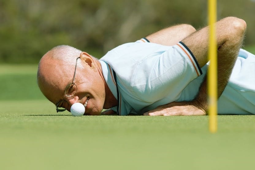
Presbyopia is usually first noticed around the age of 40 to 45 years. Although difficulties with close work may seem to come on suddenly, the aging process that causes presbyopia is gradual and has been going on since childhood. Between the ages of 45 and 65, the amount of presbyopia increases, making near work more difficult.


Presbyopia can be corrected by spectacles and/or contact lenses. It is important that the prescription is calculated for the distance at which you do your close tasks. Surgery can now correct for presbyopia through use of multifocal intra-ocular lenses and monovision, however there are associated risks and outcomes are not always ideal. A thorough discussion with your optometrist will enable you to decide on your best options.


Reading glasses, the primary correction for presbyopia will make near objects clear but distant objects blurry. This means that if you have a pair of spectacles just for reading, you will not he able to watch television while wearing them. Having different prescriptions for distance and reading can be a nuisance, especially if you have to change spectacles all the time. Bifocals, progressive lenses and ½ eyes have all been designed to reduce this problem, however in our current digital world we find ourselves prescribing specific glasses for specific tasks rather than a one size fits all.


Although your close focusing system is not functioning as well as it used to, once presbyopia has been corrected with spectacles, you will be able to see close things as well as you always did. Presbyopia does not represent a threat to your eye health and wearing spectacles will neither accelerate nor slow the development of presbyopia.


Between the ages of 45 and 65 years, your prescription is likely to change significantly. It is sensible to have your eyes examined every two years to review your correction and your general eye health. If you experience vision problems earlier, you should make a review appointment with your optometrist. Your optometrist will advise you of the most appropriate period between consultations.

Dr Ieuan H. Rees. (Optometrist)
Buck & Todd Optometrist
Located at 103 Alfred Street, Mackay, 4740.

Mackay Optometrist | Mackay Eye Doctor Testing
Read More

Amblyopia

Amblyopia = Lazy Eye

It is perfectly natural as a parent to feel anxious and a little upset by the fact that your child has lazy eye (amblyopia), particularly if you were unaware of the problem before your child’s eye examination. You may have noticed a turned eye, favouring one eye, bumping into objects on one side, head tilt or that they don’t perform certain visual and eye-hand tasks as well as expected.

Amblyopia, or ‘lazy eye’, affects up to 4% of the population and is a condition where the message, sent from the affected eye or eyes to the brain, is of low quality, making the sight of that eye poor. A person with amblyopia will have poor vision that doesn’t fully correct simply with spectacles.

Simply stated amblyopia is not “medical.” There is no disease. There is no drug or surgery to cure it. This is a neurological condition. The part of the brain that processes detailed information from that eye (or eyes) is under-developed due to lack of stimulation. The child simply has not learned to see clearly (vision) with the eye. The only treatment is to stimulate vision development.

Common causes of this are a turned eye or a large difference in the extent of shortsightedness, long-sightedness or astigmatism (refractive error) between the person’s eyes. Rare causes of Amblyopia can include disease or injury to the nerve connecting the eye to the brain, some psychiatric conditions and excessive use of tobacco, alcohol or other drugs.

Visual development is incomplete at birth. The basic components of the visual system are present, but a child’s visual system continues to develop after birth in response to the visual environment. This development progresses rapidly in the first few years of life and can be subject to stressors which impede normal progress.

In a person with Amblyopia, the message received by the brain from each eye is different. This occurs when there is lack of clear focus, or because the eyes are pointing in different directions. The brain compensates for this difference by ignoring the message from the worse eye. If a visual system is not corrected within the first 7 to 8 years, the child may never develop the ability to process the image from the worse eye. A thorough eye examination is the only sure way of determining whether your child has Amblyopia. If you think that there may be something wrong with your child’s vision, discuss it with us.

Treatment

Treatment depends on the cause. Most commonly glasses, prisms, contact lenses and vision therapy programs are used to train the poor eye to function normally. For 300 years the better eye has been covered with a patch to encourage use of the lazy eye, however recent research indicates other mechanisms such as a combination of syntonics, binasal occlusion, refractive engineering, vision therapy and monocular fixation in a binocular field are often more effective in improving acuity and encouraging effective binocular vision. Some special or rare types of Amblyopia are treated simply by improving the person’s general health. The earlier Amblyopia is detected, the easier it is to treat.

It is important to be secure in the diagnosis and be committed to the program when you do start vision therapy. A second opinion to rule out pathological causes is never discouraged.

Related brochures

  • Hyperopia (long-sightedness)
  • Myopia (shortsightedness)
  • Astigmatism
  • Vision and school achievement
  • Parents’ guide to children’s vision

Dr Ieuan H. Rees. (Optometrist)
Buck & Todd Optometrist
Located at 103 Alfred Street, Mackay, 4740.

Mackay Optometrist | Mackay Eye Doctor Testing
Read More